A reptile that suddenly wants nothing to do with food, or that pulls back when you reach in, is often hiding a problem you cannot see. Mouth rot — veterinarians call it infectious stomatitis — is one of the most common bacterial infections in pet snakes, lizards and turtles, and its earliest stage is easy to miss precisely because reptiles mask discomfort so well.
The Early Signs Are Deliberately Subtle
At the start you are looking for tiny purplish-red pinpoint spots inside the mouth, often along the gums. That is not a dramatic symptom, and it is exactly why a weekly hands-on inspection earns its place in your routine.
- Pinpoint red spots or redness along the gum line
- Swelling of the lips, mouth or jawline
- Thick, cheesy or pus-like material along the teeth and gums
- Stringy saliva or drooling, where there was only thin clear fluid
- Refusing food, or holding the mouth slightly open
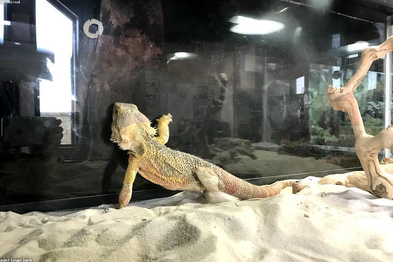
In lizards and bearded dragons, swelling tends to show first around the front of the jaw. In snakes, look for discharge along the gum line and around the labial pits.
Why It Happens: Husbandry First, Bacteria Second
The bacteria involved — Aeromonas and Pseudomonas are the usual suspects — normally live harmlessly in a reptile’s mouth. The infection takes hold when something weakens the animal or gives bacteria a way in.
The most common contributors are an incorrect temperature gradient, because reptiles rely on their environment to regulate immune function; dirty water and substrate; and physical injury from snout rubbing, striking at enclosure glass, or bites from live prey. Retained shed around the lips, vitamin A deficiency in lizards and an illness already running in the background all make the same door easier to open.
What Treatment Actually Involves
Mouth rot does not resolve on its own, and it does not respond to home first aid. Left alone it tracks along the dental arcade and can extend into the upper and lower jawbones. It matters for a second reason too: aspirating infected material can seed a secondary pneumonia. A heat lamp setup that is correct for the species is part of the fix, not an optional extra.
| Stage | Typical veterinary action |
|---|---|
| Early, localised | Oral exam, antiseptic flushing, systemic antibiotics |
| Dead tissue present | Surgical debridement under anaesthesia, repeated cleaning |
| Bone involvement | Imaging, aggressive surgery, longer antibiotic course |
| Any stage | Supportive care: fluids, assisted feeding, husbandry fixes |
Vitamin A and C supplementation is often suggested, though it does not reliably change how the disease runs. Never lance, scrape or pack a reptile’s mouth at home — the tissue is delicate and the infection spreads easily.
Preventing the Next Round
Because husbandry drives this disease, prevention is mostly environmental: hold the correct temperature gradient for your species day and night, keep water and substrate clean, and reduce stress with more hides and visual barriers so your reptile is not rubbing its snout against glass. Checking the mouth during normal handling takes seconds and is your best early warning. If you notice two or more signs at once, book the exam this week rather than waiting for the animal to look obviously sick.
General information, not a substitute for veterinary care. Reptile infections progress quickly, so early assessment matters.