Mast cells are immune cells best known for the itch and swelling of an allergic reaction, and they carry granules full of histamine. When mast cells become neoplastic, those granules explain much of the behaviour that owners see: lumps that seem to swell and shrink over days, that itch or sting, that bleed or bruise easily when handled. In cats these tumors form three separate syndromes, and where the tumor sits determines the outlook far more than how it looks.
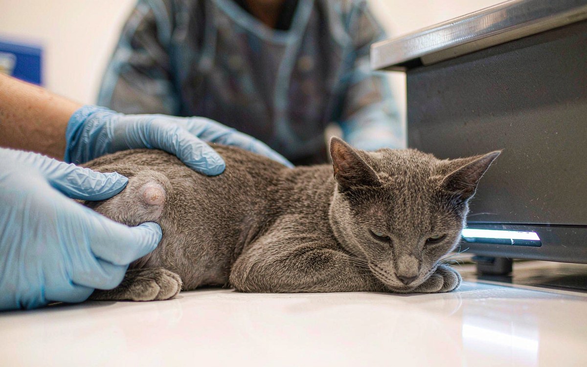
Three Forms, Three Very Different Outlooks
Cutaneous mast cell tumors sit in the skin. Visceral disease involves internal organs, most often the spleen or liver. Intestinal mast cell tumors grow in the wall of the gut. Cats differ from dogs in important ways: there is no subcutaneous category as there is in dogs, dedicated grading systems arrived later, and the skin form is far more likely to behave benignly. As a group, skin mast cell tumors are the second most common skin tumor diagnosed in cats.
What a Mast Cell Tumor Looks Like
There is no single appearance. Some are firm raised white or pink bumps within or just under the skin. Others are hairless, red, ulcerated or bruised growths that bleed when touched. Some stay the same size for months or years, others grow over days, and many fluctuate over time, which is why appearance is a poor guide. Siamese cats appear to be predisposed to the skin form, and a mutation in the KIT gene has been found in cats with these tumors.
- A lump that changes size, especially one that swells and shrinks
- A growth that itches, so the cat scratches, licks or bites at it
- An ulcerated or bleeding sore that does not heal
- New lumps appearing in groups, or later batches of lumps
- Vomiting, reduced appetite, weight loss or lethargy, which point to internal disease rather than skin
Diagnosis and Staging
A fine needle aspirate is usually all it takes to confirm a mast cell tumor, because the cells are distinctive under the microscope. From there, vets usually go further. A biopsy of the excised lump is sent to a pathologist to check the mitotic rate and the tumour’s structure, since the growth pattern of the cells is what predicts spread. Lymph nodes are sampled if they feel abnormal. Abdominal ultrasound with routine aspiration of the liver and spleen, and sometimes bone marrow sampling, check for internal involvement.
Staging is not optional. Roughly one in five cats with skin mast cell tumors has abnormal mast cells in the spleen as well. In most of those cats the findings are separate events rather than true spread, but the distinction changes what the vet recommends.
| Form | Typical behaviour | Rough outlook |
|---|---|---|
| Cutaneous, low grade | Generally benign; may recur locally if cells are left behind | Frequently cured with surgery, survival often measured in years |
| Cutaneous, high grade | More likely to spread beyond a nearby lymph node | Roughly a year on average, varying by cat |
| Splenic | Can spread to other organs | Around fourteen months with spleen removal, much shorter without |
| Intestinal | Commonly spreads to lymph nodes and liver | One to two years with aggressive treatment |
Treatment Direction
Surgery is the first choice for nearly every skin mast cell tumor, because the appearance of the lump tells you nothing about how aggressive the cells are, and a properly removed tumour may be a cure. Specimens are always sent for pathology to check whether the margins were clean. If cells were left behind, options include a second surgery, radiation therapy for tumors in awkward locations or too large to remove cleanly, or careful monitoring for regrowth. When tumors are numerous, unresectable, or have already spread, chemotherapy is used, with injectable and oral protocols available, including a tyrosine kinase inhibitor given at home as a tablet. Steroids such as prednisolone may be added to slow growth and reduce inflammation.
Two supportive details matter in cats. Because mast cells release histamine, vets often prescribe an antihistamine and an antacid together to protect the stomach from excess acid and to reduce nausea, and pain relief is planned around the procedure. After surgery, keep the incision clean and dry, use a cone or recovery suit so the cat cannot lick the site, restrict activity for the usual ten to fourteen days, and call the clinic if sutures come loose or the area swells or bleeds.
Most cats with a single low-grade skin mast cell tumor do well, and many live out a normal lifespan. Run your hands over your cat monthly, and have any new lump checked rather than watched for months.